Michael Yancey - Dr. Ting Cong - Week 6
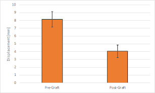
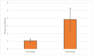
For the femoral head impaction grafting project, I began writing methods, results, and some discussion for the manuscript we intend to submit for publication. My parts of this so far include detailing the compressive loading process and subsequent mechanical analysis, such as how we calculated defect region moduli or linear displacement at 800 N, the 3D scanning process and subsequent volumetric analysis of defect volumes, and then the corresponding results. These included toe-region and linear region moduli for the fracture, linear displacement at 800N, and defect and raised volumes at various stages throughout testing—all of these also included a paired t-test analysis comparing each fracture before and after grafting.
I began generating figures for this paper, some of which are included below. In order, these show a 3D reconstruction of the Osteopearl graft beads inside the subchondral fracture volume, the linear displacement at 800N before and after grafting, and the fracture's modulus before and after grafting.

---------------------------------
For the mesenchymal stem cells in alginate hydrogels project, we created two cell-seeded gels at the beginning of the week, then images them at the end of the week. These gels were the "low molecular weight - low calcium" and "low molecular weight - high calcium" variations of the hydrogel. The gels continued to be a viable medium for cell survival as we were able to image a significant number of cells, however, cell migration was still lacking as we found only a couple cells that migrated any significant distance over the two-hour imaging session when searching manually.
---------------------------------
I also had the opportunity to attend another surgery on Wednesday. This surgery was with Dr. Joseph Lane, where the surgeons performed an avascular necrotic decompression of the femoral head. The most interesting feature of this surgery for me was its relevance to my femoral head impaction research. There are significant similarities in the femoral head after core decompression and subchondral fracture, especially in terms of the subchondral volume of space with fractured or removed bone. I was excited to see the surgeons use the Osteopearl beads that I have been researching to fill the cored volume of the femoral head. Particularly fascinating was their approach to rehydrate the beads—they used a solution of saline and the patient's own marrow to provide cells for bone remodeling during recovery.

Comments
Post a Comment